

1. Product Introduction
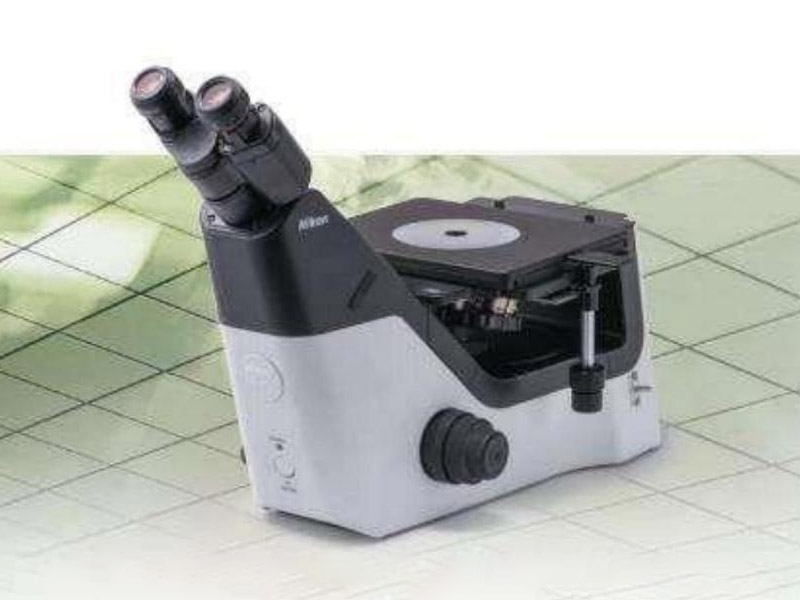
The MA-100N is a classic, cost-effective model in Nikon’s inverted metallurgical microscope product line. Adopting the CFI60-2 optical system, it delivers flat-field achromatic observation, clear imaging, high contrast, and high energy efficiency. Combining superior performance with simple operation, it provides flexible solutions for your busy material testing work.
2. Application Fields
Ferrous samples: Phase analysis; grain size, inclusion inspection, gray cast iron, nodular cast iron inspection, cemented carbide inspection.
Non-ferrous metals and alloy samples: Alloy phase analysis; grain size, inclusion inspection.
Dissection of various metal materials (before imitation): Material microstructure and heat treatment condition inspection.
Surface coating inspection: Coating thickness, surface roughness, and coating porosity measurement.
Metal welded joint inspection: Inspection of weld center zone, heat-affected zone, and base metal microstructure.
Fracture failure analysis of various metal parts.
Metallographic analysis and identification of fire accidents.
3. Basic Performance
3.1 Optical System
Adopts the standard CFI60 infinity semi-apochromatic optical system. Optical components are coated with anti-mold film, making it a genuine ISO14001 environmental-friendly product.
Magnification: 50x - 500x.
Adopts fly-eye lens technology for a more uniform and bright field of view.
Observation method: Bright field.
Ergonomic design, sturdy and stable with high load capacity, simple and comfortable operation.
High manufacturing precision with position encoding; all calibrated technical parameters are the lower limits of actual values. Pre-installation and debugging are carried out before delivery to ensure the quality and matching of all components.
3.2 Lenses
Eyepieces: Wide-field binocular observation, 10x/22 high eye-point with diopter compensation. Large and comfortable observation field of view. The binocular tube is built-in hinged type with adjustable eye-point height. The 45° tilt angle conforms to the most comfortable sitting posture. It can be equipped with a standard C-mount for high-definition photography.
Objectives: Adopts the latest CFI60-2 series objectives. Based on the high numerical aperture and long working distance realized by the CFI60 optical system, it features longer working distance, better achromatic aberration, improved low-magnification performance, and lightweight design. Specifications: 5x, 10x, 20x, 50x.
3.3 Mechanical Parts
Mechanical Stage: Multiple stages are available with sturdy and stable specimen clamps. The MA-SR rectangular stage with three-plate mechanism is selected, which can hold large and heavy specimens with a large moving range. X-axis and Y-axis travel: 50x50mm, with coaxial control handle on the right side.
Nosepiece: 5-hole bright field objective turret with high precision between positions, ensuring better parfocality.
Focusing: Coaxial coarse/fine focusing knobs with 8.5mm travel (37.7mm stage movement per full rotation of coarse knob, 0.2mm stage movement per full rotation of fine knob). Built-in Nikon high-precision rack and pinion, preventing automatic sliding during long-term use.
3.4 Imaging Device
Equipped with a professional digital high-speed camera (industrial camera) with 5 million effective pixels.
3.5 Light Source: LED Light Source
4. Image Analysis System
4.1 Hardware
DELL commercial computer, HP printer.
4.2 Software
Professional metallographic analysis software (Chinese version) sets a new standard in image processing, analysis, and file management. Featuring a unique and harmonious modular design, it meets basic digital image requirements and can be extended for complex advanced applications in material microstructure research.
Developed based on national inspection standards, the software includes more than 100 functional modules. Users can select inspection items as needed and complete inspections with the software’s assistance.
Functions include:
Automatic rating: The software automatically generates final analysis results.
Assisted rating: The software provides parameters related to the analysis results.
Comparative rating: Compares captured specimen images with standard image libraries for manual judgment.
4.3 Working Principle
Cameras convert optical images from the microscope into digital signals and input specimen images into a computer. Professional image analysis software processes and measures the digital images. Finally, images and measurement results are printed and output, and can be backed up to a CD using a CD burner.
4.4 Image Analysis and Measurement
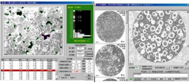
Initial magnification calibration with accuracy < ±5%. Provides various measurement parameters: point, line, area, angle measurement, target area, perimeter, major/minor diameter, shape factor, average gray level, absolute optical density, nucleocytoplasmic ratio, etc. Measurement results can be displayed in data lists, histograms, site maps, etc. The system automatically segments, counts, statistics, classifies, and measures target images, assigns serial numbers, and displays parameters for each measured target.
4.5 Image Processing
Adjust image color, brightness, and contrast; binarization for grain size display; filter, sharpen, soften, edge enhancement; image segmentation, stitching, and overlay.
4.6 Automatic User Report Generation
Users can record each operation step to create a processing program. Inspection reports are generated automatically with one mouse click.
(Image Example)
5. Environmental Requirements
5.1 Operating Environment
Anti-vibration: Keep away from vibration sources.
Moisture-proof: Use air conditioner or dehumidifier.
Dust-proof: Equip with anti-static plastic flooring and dust-proof walls.
Temperature: 20℃±10℃
Humidity: Relative humidity ≤ 85%
Curtains: Double-layered, inner red-black, outer light-colored.
5.2 Power Supply: 220V±10%, 50Hz
5.3 Workbench
Size: ≥ 70cm×140cm
Tabletop: Non-flammable material.
6. Training
Training Content: Operation of the metallographic microscope and imaging system, basic maintenance of the microscope, software operation.
Training Location: Customer’s site.
Trainees: 2 persons.
Training Duration: 1 working day, enabling operators to work independently.
7. After-Sales Service
7.1 The buyer shall provide detailed fault information when equipment failure occurs. The seller shall respond within 8 hours upon receiving fault information, including verbal guidance for troubleshooting.
7.2 If the fault cannot be resolved remotely, the seller’s personnel will arrive at the site to solve the problem within 48 hours.
7.3 The seller provides free software upgrades during the warranty period.
8. Warranty
12-month free warranty after commissioning and acceptance by the buyer (except for man-made damage).
9. Scope of Supply
| No. | Item | Quantity |
|---|---|---|
| 1 | ECLIPSE MA100N Main Body | 1 |
| 2 | TS2-W 10x Eyepiece Lens | 2 |
| 3 | MA-SR-N Rectangular Stage N | 1 |
| 4 | MA-SRSH1 Universal Specimen Holder | 1 |
| 5 | MTI LE PLAN EPI 5x (NA/WD: 0.10/31mm) | 1 |
| 6 | MTI LE PLAN EPI 10x (NA/WD: 0.25/13mm) | 1 |
| 7 | MTI LE PLAN EPI 20x (NA/WD: 0.40/3.6mm) | 1 |
| 8 | MTI LE PLAN EPI 50x (NA/WD: 0.75/0.50mm) | 1 |
| 9 | TS2-P-CF Camera Port 100 Beam Splitter | 1 |
| 10 | Metallographic Analysis Software | 1 |
| 11 | 9MP Industrial Camera | 1 |
| 12 | Branded Commercial Computer | 1 |
| 13 | HP Inkjet Printer | 1 |